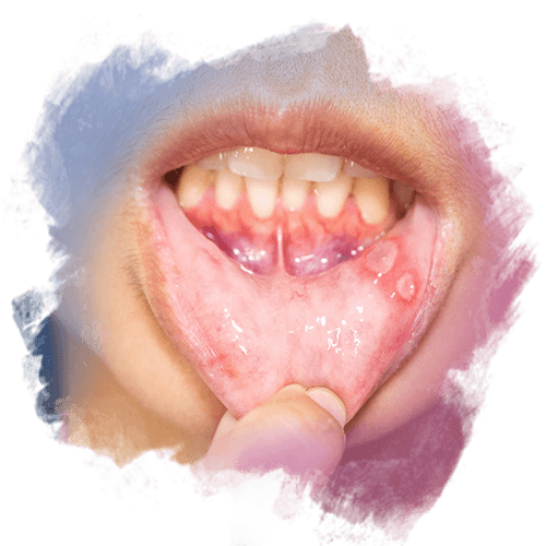
Gum discoloration is a condition characterized by the gingival tissue changing from its normal pink tone to a purple, dark red, or bluish appearance. While it creates aesthetic concerns, it can also be an indicator of periodontal health. This color change may develop due to increased melanin pigmentation, vascular changes, trauma, inflammation, smoking, tissue reactions around implants, medication effects, or genetic pigmentation. Not every discoloration is pathological, but the surrounding tissue health, blood supply, and tissue nutrition must be evaluated. The goal is not only to lighten the color, but to identify the underlying cause and support tissue biology.

Diş Eti Morarması Nedir? Nasıl Açılır?

[Total: 0 Average: 0]

Main Causes of Gum Discoloration

Melanin Pigmentation

In some individuals, naturally high melanin production is observed. Especially in people with darker skin tones, the gums may appear darker, and this is considered physiological.

Smoking

Nicotine and toxins in tobacco cause vasoconstriction and pigment changes in the gingival vessels. This often results in a dark purple to black appearance.

Trauma and Inflammation

Gum pressure, trauma, or previous periodontal diseases can cause tissue damage and impaired blood circulation, leading to discoloration.

Medication Effects

Some medications (such as antimalarial agents, minocycline, etc.) may cause gum darkening as a side effect due to pigment deposition.

Metal Implant Abutment Visibility

In metal-based implant restorations instead of zirconium, metal shine-through—especially in thin gingival biotypes—can lead to a purple-gray appearance of the gums.

Vascular Structure and Hematomas

Small vascular enlargements in the gums or post-traumatic hematomas can also leave purple-like spots or discoloration.

How to Lighten Gum Color?

Laser Depigmentation

A laser is applied to the pigmented area, and melanin cells are selectively removed, allowing the tissue to achieve a more pink and natural appearance. One session may be sufficient; in some patients, repeated treatments may be planned.

Surgical Depigmentation

The superficial gingival layer is gently removed, allowing new epithelial tissue to develop. It is an effective option in individuals with high melanin density.

Periodontal Treatment and Inflammation Control

If discoloration is due to inflammation, professional cleaning, scaling, root surface debridement, and a home care protocol are required.

Smoking Cessation

As the vascular structure recovers, the tissue color gradually returns to a healthier pink tone.

Implant-Related Soft Tissue Management

If the dark appearance is caused by an implant, the following can improve tissue color:

  • Zirconium abutment
  • Connective tissue graft
  • Gingival thickening

Single-Item Clinical Approach

Pigment Source + Tissue Biotype + Technique Combination

  • Pigment source analysis
  • Melanin density mapping
  • Biotype evaluation (thin/short/thick)
  • Selection of laser or surgical depigmentation
  • Periodontal inflammation control
  • Peri-implant tissue thickening
  • Home care and smoking support plan
  • Planning a second session when necessary

This approach is not only about lightening the color;
it aims for tissue health, aesthetic contour, and a natural gingival tone.

Although gum discoloration may seem like a purely cosmetic issue, pigment biology, vascular structure, tissue thickness, and periodontal health should all be evaluated together. While laser and surgical depigmentation techniques provide an aesthetic pink tissue appearance, smoking cessation, inflammation management, and peri-implant tissue support also offer long-term results. With proper planning, the gingival tone can return to a healthier, brighter, and more natural pink gingival aesthetic form.

Leave a Reply

Your email address will not be published. Required fields are marked *

This is a sample text. You can click on it to edit it inline or open the element options to access additional options for this element.